Introduction: You’ve Done This Dozens of Times and Never Really Felt It

Eye drops in, head tilted back, trying not to blink. A few seconds of mild effort, then release — and it’s over before it registered.
But those few seconds are neurologically dense. The most sensitive tissue in the body is receiving a chemical stimulus. The brain’s automatic blink control is being manually overridden. A specific tension is building, and then it’s released.
What feels like a minor routine is arriving at tissue that is, neurologically, extremely well-equipped to receive it. The whole cycle takes about thirty seconds. Today’s practice is about being present for all of it.
Session 1: Why This Particular Moment

The cornea is among the most densely innervated tissues in the human body — estimated at roughly 300 to 600 times the sensory nerve density of skin. Branches of the trigeminal nerve’s ophthalmic division distribute throughout the corneal epithelium and stroma, providing high-sensitivity detection of temperature, mechanical stimulation, and chemical input. This density is evolutionary design: the eye requires rapid, precise threat detection at its outermost surface.
The result is that eye drops don’t feel like anything else. The coolness, the spread of liquid across the corneal surface, the immediate sensory response — these are inputs to one of the body’s most sensitive receptor fields. What registers as a minor everyday sensation is arriving at tissue that is, neurologically, extremely well-equipped to receive it.
The second layer is the blink itself — or rather, the suppression of it. Blinking at rest is an automatic rhythmic motor behavior maintained by the basal ganglia and supplementary motor area, cycling every three to five seconds without conscious direction. To consciously hold it back requires descending inhibitory signals from the prefrontal cortex to interrupt that automatic cycle. For the duration of the suppression, automatic motor control and conscious control are running simultaneously in opposition. That subtle tension while waiting to blink is this opposition made physical.
Session 2: Three Steps
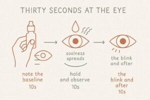
STEP 1: Start when you pick up the bottle (10 seconds)
The practice begins before the drops go in. With the bottle in hand, check the current state of the eyes — tired, dry, strained, or simply there. Whatever is present, note it. This is the baseline the practice will compare against.
STEP 2: While holding the blink (10 seconds)
From the moment the drops make contact, open the sensory channels.
The corneal sensation — the temperature of the liquid, the quality of the coolness, the way it spreads
The suppression itself — the subtle tension of the eyelids held open, and the impulse to blink arriving in waves, not steadily
The breath — the tendency to hold it; let it continue
Observe without rushing toward the release.
STEP 3: The blink and after (10 seconds)
When the blink is allowed — follow what happens. Where does the tension release from first? How does the liquid redistribute? When does the breath return? How does the visual field change in the seconds immediately after?
The cycle completes. Stay with it through the end.
Session 3: What’s Actually Happening in the Thirty Seconds You’re Trying Not to Blink
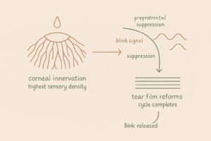
Corneal innervation density is among the highest of any tissue in the body, with estimates ranging from 300 to 600 times that of skin. The ophthalmic branch of the trigeminal nerve sends sensory fibers throughout the corneal stroma and epithelial layers, providing dense coverage for thermal, mechanical, and chemical detection. This architecture exists because the cornea is the eye’s first line of contact with the external environment — evolutionary pressure has made it exceptionally sensitive to anything that touches it. A liquid at a different temperature than the tear film, spreading across a receptor field this dense, produces an input that is qualitatively unlike most other daily sensory events.
The blink suppression introduces a separate mechanism. Spontaneous blinking is a rhythmic automatic behavior regulated by the basal ganglia and supplementary motor area — part of the same automatic motor circuitry that maintains gait, posture, and other background motor behaviors. The system runs continuously without requiring attention. To consciously suppress it requires the prefrontal cortex to generate descending inhibitory signals that interrupt the supplementary motor area’s automatic cycle. For the duration of the suppression, two systems are active and in opposition: the automatic system generating blink commands, and the prefrontal system blocking their execution.
The impulse to blink that builds during suppression is not a single sustained pressure. It arrives in waves — reflecting the automatic system repeatedly generating the blink signal and the prefrontal system repeatedly suppressing it. Observing the impulse as waves rather than as uniform tension is an invitation to watch this back-and-forth directly. This is distinct from emotional or cognitive impulses — what’s being observed here is a pure motor control signal, the basal ganglia’s automatic output, visible to awareness only because the prefrontal cortex is actively blocking it.
When the prefrontal suppression is lifted, the automatic system executes the blink: the orbicularis oculi contracts, the tear film is redistributed across the corneal surface, and the optical uniformity of the surface is restored. The clarity of vision in the seconds immediately after blinking — the specific quality of a just-refreshed visual field — is the direct perceptual correlate of tear film reformation. The corneal surface is optically smoother. The image is cleaner. Receiving this consciously, rather than simply moving on, is the completion of a sensory cycle that the body ran from beginning to end in about thirty seconds.
Conclusion: The Nervous System Was Busy the Whole Time

Once today. One application, from picking up the bottle to the blink and after — the whole cycle, nothing skipped. The drops last three seconds. Everything around them takes the other twenty-seven.
The tissue was always this sensitive. Most days, nothing asks it to prove that.
KEY TERMS
Corneal Innervation Density
The cornea carries an estimated 300–600 times the sensory nerve density of skin, via branches of the trigeminal nerve’s ophthalmic division. Evolutionary design for rapid threat detection at the eye’s outermost surface. The neuroanatomical reason eye drops produce a qualitatively distinct sensory experience — the input is arriving at one of the body’s most sensitive receptor fields.
Neural Control of Blink Suppression
Spontaneous blinking is maintained by the basal ganglia and supplementary motor area as an automatic rhythmic motor behavior. Conscious suppression requires descending inhibitory signals from the prefrontal cortex to interrupt this automatic cycle. The subtle tension of holding the blink is the felt experience of these two systems running in opposition simultaneously.
The Blink Impulse as Motor Signal
The impulse to blink during suppression arrives in waves — the automatic system generating the blink command repeatedly, the prefrontal system suppressing it repeatedly. A pure motor control signal made visible to awareness only because it’s being blocked. Distinct from emotional or cognitive impulses — this is the basal ganglia’s automatic output, observable precisely because the prefrontal cortex is actively interrupting it.
Tear Film Reformation
The redistribution of tear fluid across the corneal surface that occurs with each blink, restoring optical uniformity. The improved visual clarity in the seconds after blinking is the direct perceptual experience of this reformation — the corneal surface smoother, the image cleaner.
Defusion
A core skill in Acceptance and Commitment Therapy (ACT): the capacity to observe thoughts and impulses as passing mental events rather than directives. When just get this over with arrives during the suppression, recognizing it as a thought rather than a command — and staying with the corneal sensation for the remaining seconds — is defusion applied to one of its smallest possible scales.